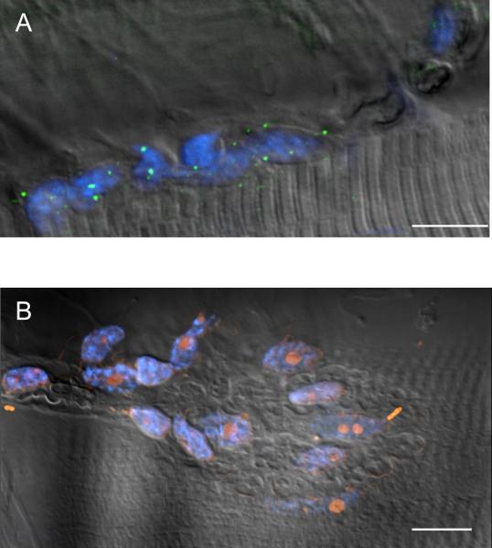
Figure 3. Immunohistochemical staining of NAAG hydrolase enzyme, GCPII.
A. Perisynaptic Schwann Cell (PSC) unablated preparation shows GCPII (green) stain at the nerve terminal (DAPI in blue indicating PSC nuclei). (N=4, n=100). B. Ablated preparation lacks GCPII (green) where ablated PSCs are marked with EthD-1 (red) and DAPI (blue) (N=2, n=50). For both panels A and B, an image collected using DIC optics is superimposed onto fluorescent images collected using DAPI, FITC and TRITC filter cubes. Calibration bars, 10 μm.

