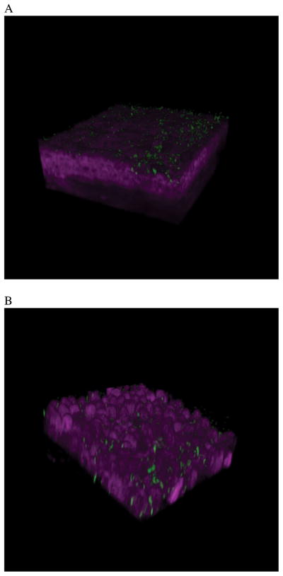
Figure 3.
Two-photon microscopy was used to show P. aeruginosa adhesion and then traversal of a MyD88 (−/−) mouse cornea at 4 h (A) and 8 h (B) post-inoculation. The cornea was not blotted with tissue paper prior to bacterial inoculation. However, bacteria readily attach after 4 h, and show extensive epithelial traversal after 8 h. A normal cornea does not show P. aeruginosa attachment or traversal. These data suggest that TLR and IL-1R signaling is significant for defending the cornea against microbial challenge. 21 (Modified from Tam et al. PLoS ONE. 2011; 6(8): e24008).

