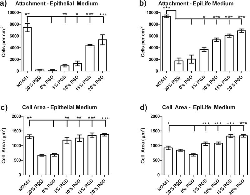
Figure 2. HCEC attachment and cell area increased with increasing levels of RGD.
HCEC attachment in epithelial medium (a) and EpiLife® medium (b) monotonically increased with increasing %RGD in the RGD/d-glucamine solution used to functionalize topographically patterned and flat substrates. HCEC projected area in Epithelial (c) and EpiLife® (d) media also demonstrated a monotonic increase with increasing %RGD. Since cell attachment and spreading was equal on topographically patterned and flat substrates, the data presented here is representative data over all of the surfaces. NOA81 substrates, which allow for non-specific protein adsorption, were used as a positive control for cell attachment and spreading. *0.01≤P<0.05, **0.001≤P<0.01, ***P<0.001

