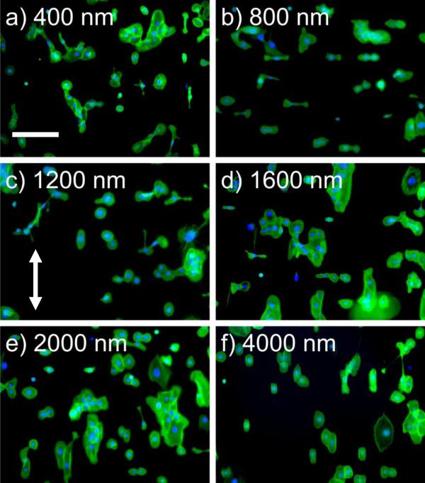
Figure 4. The presence of RGD does not promote a change in the HCEC contact guidance in Epithelial medium.
HCECs in epithelial medium on functionalized (20%RGD/D-glucamine) topographically patterned surfaces demonstrate a similar alignment response. Representative images of HCEC cells on topographic 20% RGD/d-glucamine-modified surfaces stained with FITC-phalloidin (green) and DAPI (blue). The arrow denotes the direction of the ridge/groove structures. Predominantly parallel alignment was observed for HCECs on a) 400 nm and f) 4000 nm pitch ridge/groove features. Strong perpendicular alignment was observed for HCECs on b) 800 nm pitch topography. A mixture of parallel and perpendicular cells were observed on c) 1200 nm, d) 1600 nm and e) 2000 nm pitch topography. Scale bar = 200 μm.

