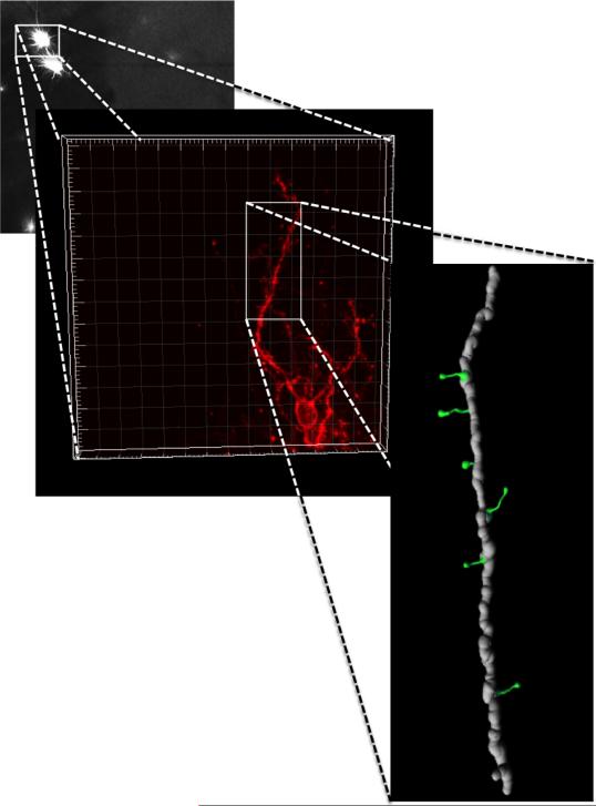
Figure 4. Three-dimensional reconstruction of confocal images.
DiI labeled medium spiny neurons in NAc were imaged and DiI was excited using the Helium/Neon 543 nm laser line. Each neuron was scanned at 0.15μm intervals along the z-axis (maximum 90 planes, depending on the depth of whole neuron and objective WD), and the morphology of the dendritic tree was reconstructed in 3-D for analysis with IMARIS software.

