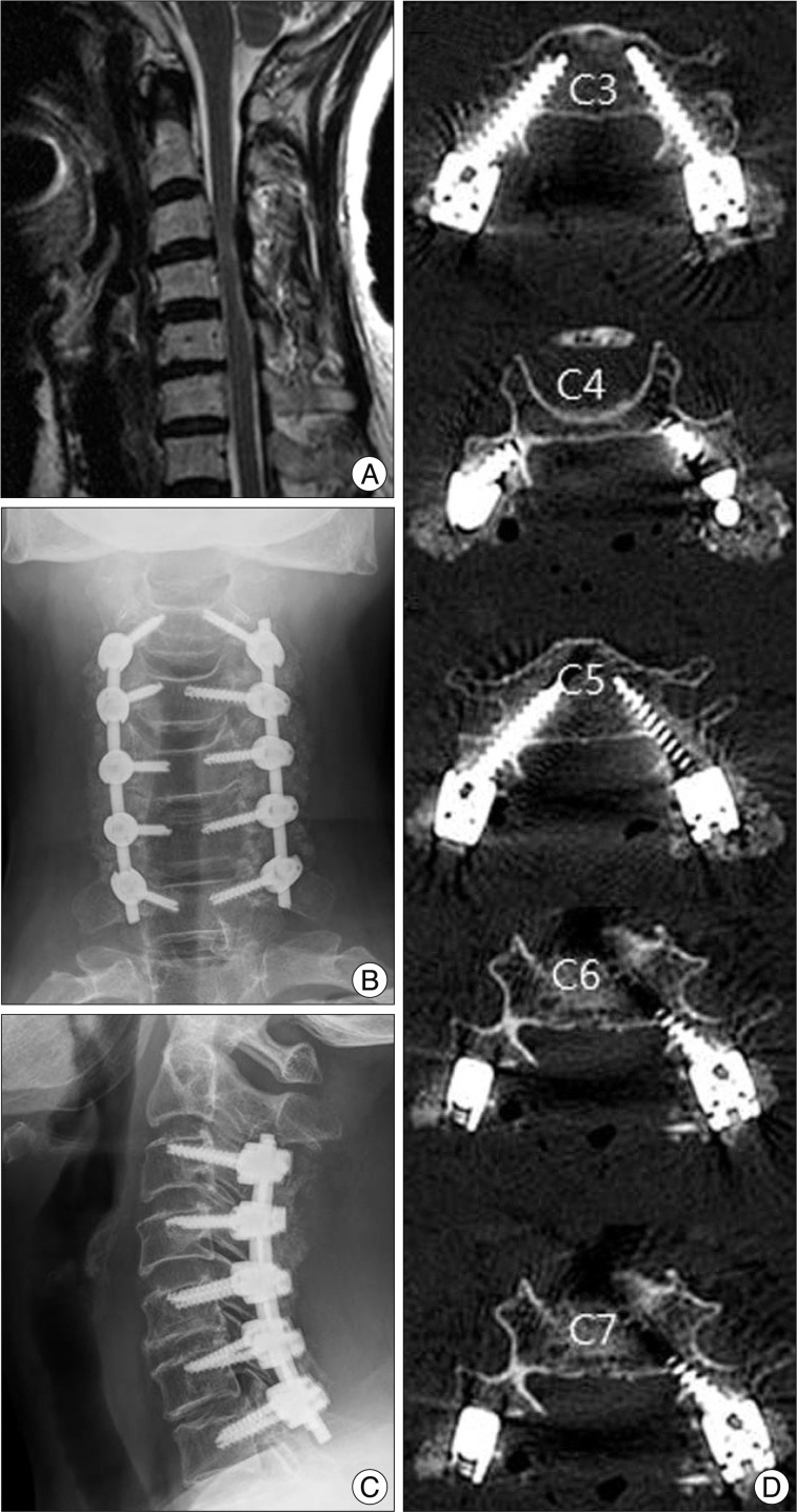
Fig. 5.
Preoperative sagittal MRI image (A) of a 71-year-old female myelopathic patient showing severe cervical cord compression. Posterior decompression with pedicle screw fixation from C3 to C7 was performed (B and C). Postoperative axial CT images show all grade 0 screw position without pedicle perforation (D).

