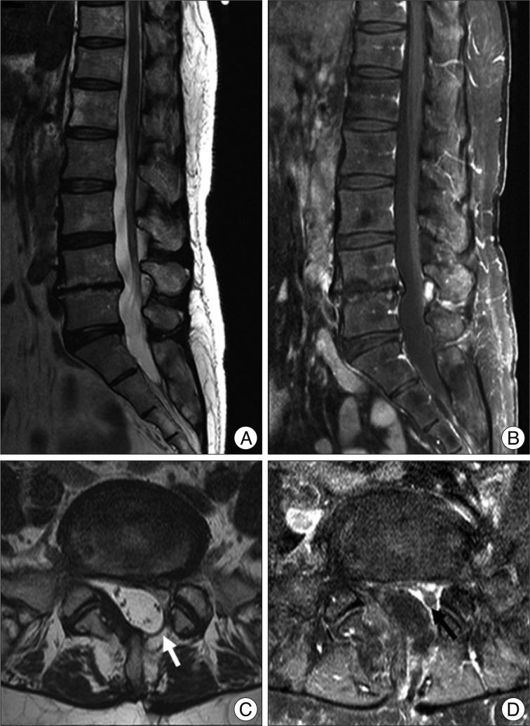
Fig. 1.
Lumbar sagittal T2 weighted (A), Gd-enhanced T1 weighted (B), axial T2 weighted (C), and Gd-enhanced T1 weighted (D) magnetic resonance imaging scans showing the postoperative changes of L4-5 level, which presenting no recurrence of disc herniation except for the findings of mild adhesion and left partial hemilaminectomy of L4 (white and black arrows).

