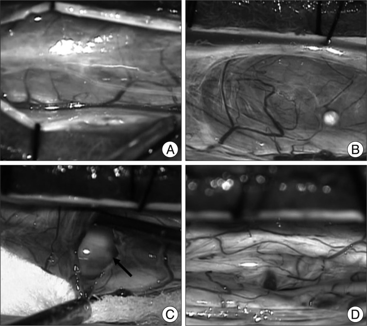
Fig. 5.
Intraoperative photographs. A : After hemilaminectomy, the dura mater is opened. B : A large arachnoid cyst, which presents well demarcated by thin and transparent wall and contains cerebrospinal fluid, is observed in the dorsal part of the intradural space, compressing and displacing the cauda equina anteriorly. C : Cystic wall and enlargement of the window were performed. No connection of the cyst with subarachnoid space is noticed. D : After partial removal of cyst wall, nerve roots are exposed and decompressed.

