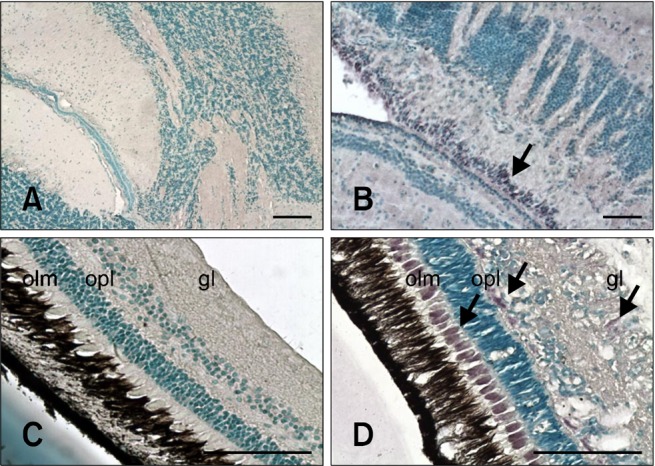
Fig. 2.

Sections of nervous tissues evaluated with IHC. Positive cells contain dark purple signals (arrows). (A) Brain from non-infected fish (negative control). (B) Brain from inoculated fish 24 days PI. Staining is in the area between the tectum opticum and cerebellum. (C) Retina from non-infected fish (negative control). (D) Retina from inoculated fish 17 days PI. Stained cells are in the outer limiting membrane (olm), outer plexiform layer (opl), and ganglion layer (gl). Scale bars = 100 µm.
