Fig. 3.

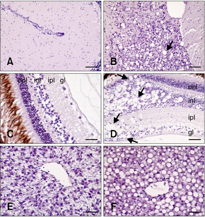
Histological damage caused by RGNNV infection. (A) Brain from non-infected fish (negative control). (B) Brain from infected fish. Vacuolation was observed in the cerebellum (arrow). (C) Retina from non-infected fish (negative control). (D) Retina from infected fish. Vacuoles (arrows) were found in the outer nuclear layer (onl), inner nuclear layer (inl), inner plexiform layer (ipl), and ganglion layer (gl). (E) Liver from non-infected fish (negative control). (F) Accumulation of fat vacuoles in hepatocytes from challenged fish. All tissues shown were sampled 17 days PI. H&E stain. Scale bars = 50 µm (B, C, and D), and 25 µm (A, E, and F).
