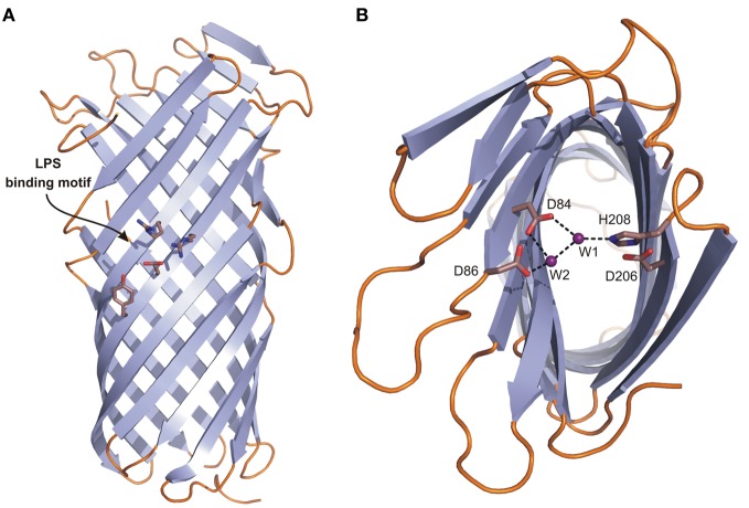
Figure 5.
The crystal structure of Pla from Y. pestis. Topology diagrams of the Pla structure (2X55). (A) Side view presenting the position of the putative LPS binding site (residues D84, D86, D206, and H208 in stick representation). (B) Top view showing the active site. D84 and D86 coordinate nucleophilic water molecule W1 either directly or indirectly via second water molecule W2. D206 and H208, the latter being hydrogen bonded to W1, form a catalytic dyad.

