Fig. 1.
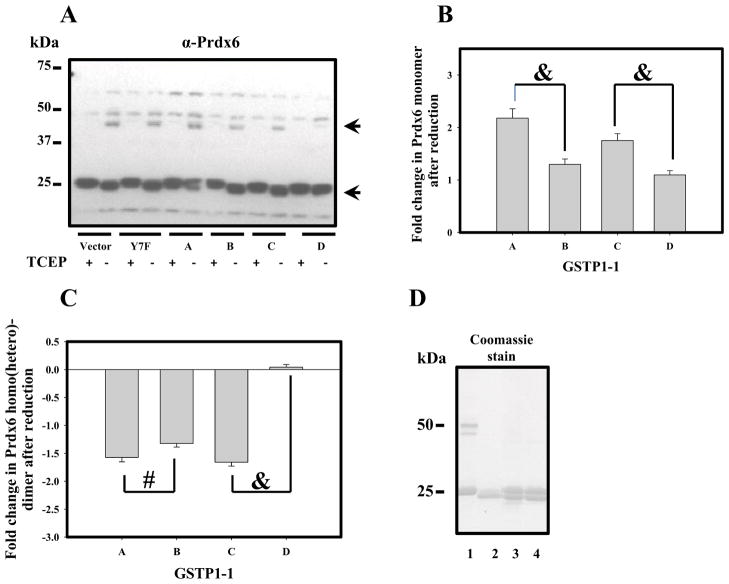
Prdx6 detection and redox state evaluation in WT and GSTP1-1-trasfected MCF-7 cells. Panel A: Immunoblot detection of Prdx6 in MCF-7 cell lysates (50μg total protein) under oxidized or reduced (2mM TCEP) conditions. Upper arrow indicates homo/hetero-dimer and lower arrow - monomeric Prdx6. Panels B and C: quantification of Prdx6 monomer (panel A, lower arrow) and of Prdx6 homo/hetero-dimer (panel A, upper arrow) content before and after reduction with TCEP. Data represent MEAN±SD for 3 independent experiments (“&” represents p≤ 0.001, and # represents p≤ 0.05). Panel D: SDS PAGE of: commercial Prdx6 (lane 1, 2μg); E. Coli expressed and purified GSTP1-1A (lane 2, 2μg), and Prdx6 mixtures with E.Coli expressed and purified GSTP1-1B or 1-1C (lanes 3 and 4, respectively; 2μg of each protein).

