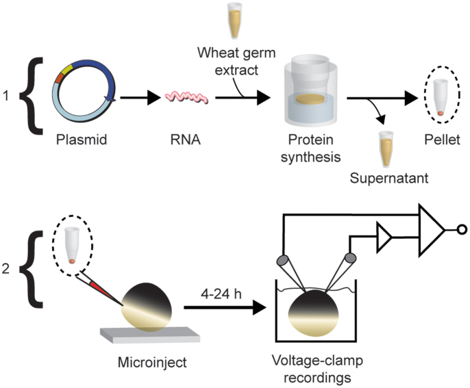
Figure 1. Cartoon workflow diagram of cell-free protein production and expression in Xenopus laevis oocytes illustrated in two phases.
Phase 1: Unique 5′ and 3′ restriction sites appended onto a target gene open-reading frame are used to digest and subclone into a specialized plasmid (pEU-HSBC) for cell-free synthesis. The plasmid DNA is used as a template for in vitro transcription of RNA. Transcribed messenger RNA directs protein translation in wheat-germ cell-free extract supplemented with liposomes to make proteoliposomes. Since the Shaker channels can be recovered from the pellet fraction, the supernatant is discarded and the proteoliposomes are collected after centrifugation. Phase 2: Samples are reconstituted to specified concentrations in a salt buffer and injected into single Xenopus laevis oocytes at the vegetal equator. Currents are measured under voltage-clamp within 24 h.

