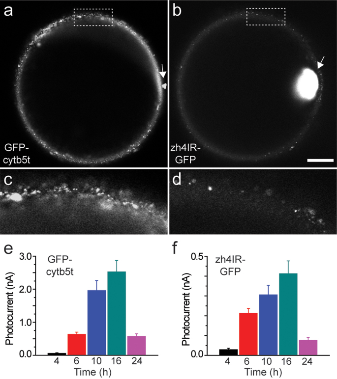
Figure 4. Membrane localization of GFP fused proteins injected into Xenopus laevis oocytes.
Grayscale confocal images of (a) GFP-cytb5t (at 2.77 pmol) and (b) zh4IR-GFP (at 0.58 pmol) protein localization within the oocyte membrane at 12 h after proteoliposome injections. Image intensity of the zh4IR-injected oocytes is increased 2-fold for ease of visualization. Arrows indicate the site of injection. Magnified membrane sections taken from the region within the dotted rectangle are displayed below. Scale bar = 200 μm (a, b) and 50 μm (magnified regions, c, d). Corresponding membrane photocurrents (nA) were measured from GFP-cytb5t (e) and zh4IR-GFP (f) at multiple times post-injection.

