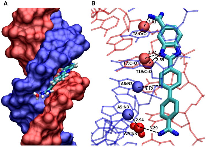
Figure 7.
The figure is from a crystal structure by Neidle and coworkers (2B0K. pdb). (A) The DNA duplex is shown in space filling with one strand in blue and one in red. DB921 is shown as large tubes in the AATT minor groove sequence. The phenyl-amidine that rises off the floor of the groove points towards the reader in this view. The interfacial water that connects this end of the DB921 complex to the floor of the groove can be seen near the floor of the groove with important DNA contact atoms in space filling representation, T-C=O in red and AN3 in blue. H-bonds are shown as dashed lines. (B) The contacts that help with the strong affinity of DB921 are highlighted. Starting at the phenyl-amidine end (bottom of the diagram) there is the interfacial water-AN3 H-bond (this is the first A of AATT) and the water is closely H-bonded to other water molecules in the minor groove. The phenyl proton that is meta to the amidine points into the groove and makes a close contact with the interfacial water. The central phenyl has a close –CH ••AN3 contact that certainly provides a stabilizing interaction. The benzimidazole –NH points into the center of the groove and forms a bifurcated H-bond with the two middle T C2=O of AATT. The benzimidazole amidine –NH that points into the groove forms an H-bond with the last T C2=O of AATT. These contacts as well as the van der Waals contacts with the floor and walls of the groove and the amidine positive charges, result in a very high binding for DB921 binding to the AATT sequence. These features could not be seen without the structural model.

