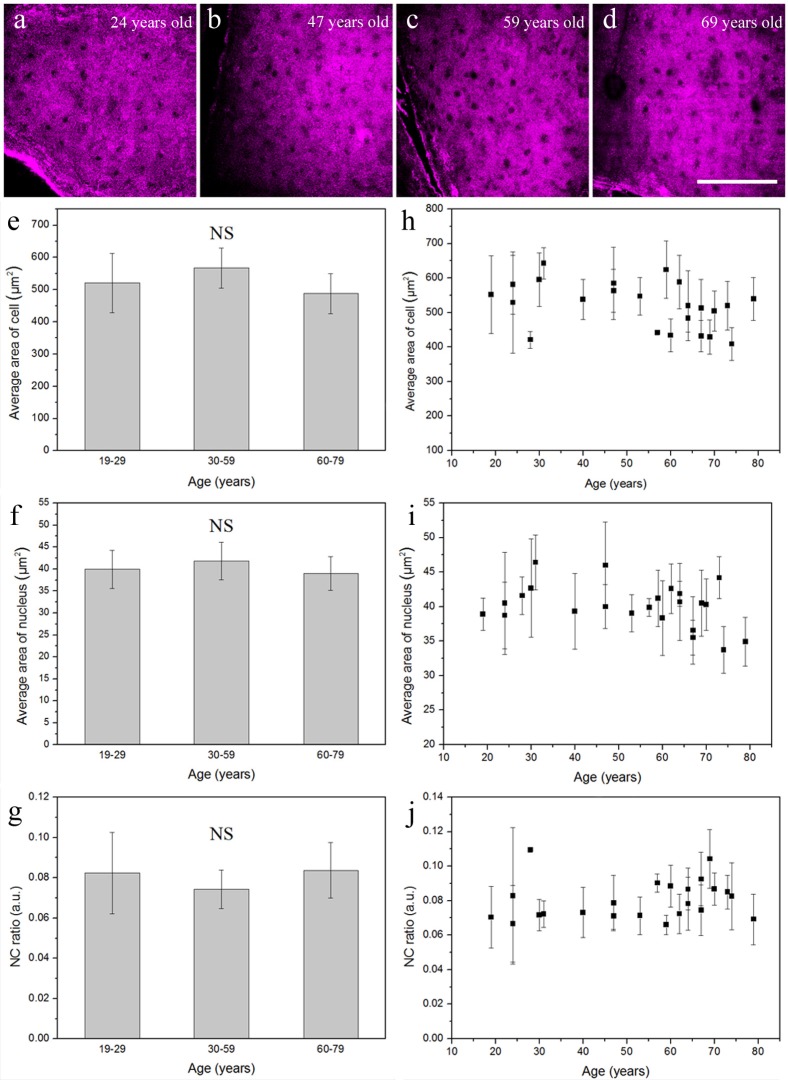
Fig. 3.
Four representative in vivo THG images of epidermal granular cells obtained from the volar forearm of (a) 24-, (b) 47-, (c) 59-, and (d) 69-year-old volunteers. In vivo cytological analysis of (e) cellular size, (f) nuclear size, and (g) NC ratio of the granular cells showed no statistical difference between these three age groups. There was no significant correlation between (h) age and cellular size, (i) age and nuclear size, or (j) age and NC ratio. NS, no significance. Scale bar = 100 μm.

