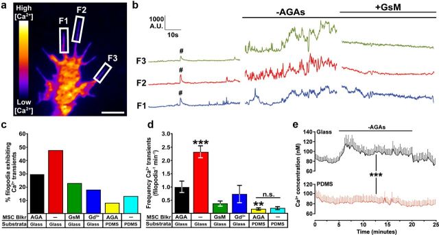
Figure 2.
MS channel activity increases the incidence and frequency of filopodial Ca2+ transients and baseline [Ca2+]i. a, A pseudocolored Fluo-4-loaded growth cone showing regions used to measure fluorescent intensities over time. Images were captured at 10 Hz for 3 min during solution changes. Scale bar, 3 μm. b, Traces of Fluo-4 fluorescent signals measured in three filopodia over 1 min periods before and after the removal of AGAs and 1 min after the addition of GsMTx4. #, denotes a single global Ca2+ transient. c, d, The incidence (c) and frequency (d) of filopodial Ca2+ transients were determined (see Materials and Methods) in control media (n = 290), in the absence of AGAs (n = 237), and after the addition of GsMTx4 (n = 174) or Gd3+ (n = 159) on FN-glass substrata. Filopodial Ca2+ transients were also measured on flexible FN-PDMS with and without AGAs (n = 103 and 52, respectively). **p < 0.01 and ***p < 0.001 compared with control condition (+AGA on glass) using a Kruskal–Wallis test with a Dunn's post test. e, Fura-2, a ratiometric Ca2+ indicator, was used to determine the [Ca2+]i within growth cones during AGA subtraction. There was a significant increase in the baseline [Ca2+]i of growth cones on FN glass after the removal of AGAs, but not by growth cones on FN-PDMS (***p < 0.001, two-way ANOVA).

