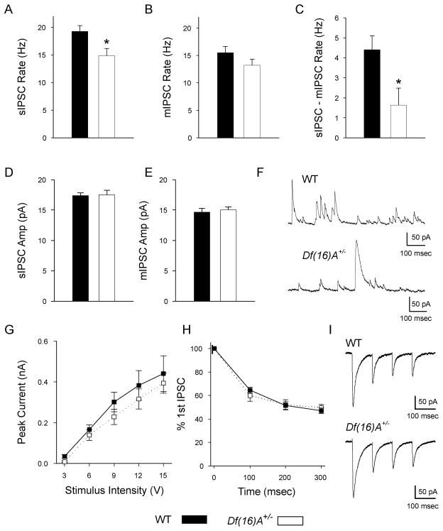
Fig. 3. Inhibitory synaptic input to CA1 pyramidal neurons.
(A) The frequency of sIPSCs was 22.9% significantly lower in Df(16)A+/− neurons (14.9 ± 1.3 Hz, n = 15) than in WT neurons (19.3 ± 1.1 Hz, n = 17, p = 0.01). (B) WT and Df(16)A+/− neurons had comparable rates of mIPSCs (WT: 15.5 ± 1.2 Hz, n = 14; Df(16)A+/−: 13.2 ± 1.1 Hz, n = 15; p = 0.16). (C) The number of events attributable to firing of INs was reduced by 62.9% in Df(16)A+/− neurons (WT: 4.4 ± 0.7 Hz, n = 14; Df(16)A+/−: 1.6 ± 0.9 Hz, n = 15; p = 0.02). (D) The average amplitude of sIPSCs were similar in both genotypes (WT: 17.4 ± 0.4 pA, n = 17; Df(16)A+/−: 17.5 ± 0.7 pA, n = 15; p = 0.88). (E) The average amplitude of mIPSCs were also similar in both genotypes (WT: 14.6 ± 0.7 pA, n = 14; Df(16)A+/−: 15.0 ± 0.5 pA, n = 15; p = 0.61). (F) Example traces from a WT neuron, top, and a Df(16)A+/− neuron, bottom, showing sIPSCs recorded at 0 mV. (G) eIPSCs from stimulation of the SR region above the pyramidal neuron were not significantly different over a range of stimulus intensities (2-way, repeated measures ANOVA, genotype: p = 0.47, genotype × stimulus intensity: p = 0.94, WT, n = 10; Df(16)A+/−, n = 6). (H) Stimulation of GABAergic axons 4 times at 10 Hz showed no difference between short-term dynamics in WT and Df(16)A+/− neurons (2-way, repeated measures ANOVA, genotype: p = 0.90, genotype × stimulus number: p = 0.32, WT, n = 10; Df(16)A+/−, n = 6). (I) Example traces from a WT neuron, top, and a Df(16)A+/− neuron, bottom, of eIPSCs recorded at −90 mV in AP-V and NBQX.

