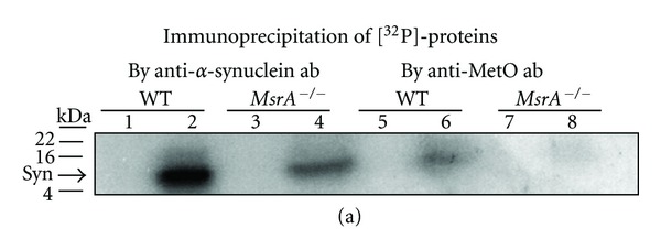
Figure 2.

Phosphorylation of α-synuclein in MsrA −/− and wild-type (WT) brain extracts. (a) Tris-soluble and Urea-soluble brain extracts (40 μg protein) of both mouse types were prepared as described in Section 2. These extracts were then incubated in the presence of additional brain-matched Tris-soluble extract (10 μg protein, serving as a source for kinases), 25 mM Tris (pH 7.4), protease inhibitor cocktail (no-EDTA) (Roche), 1 mM CaCl2, 10 mM MgCl2, and 16.7 μM [γ-32P]-ATP for 3 minutes at room temperature in a final volume of 50 μL. Endogenous phosphorylation was stopped by addition of 10 mM EDTA, 10 mM EGTA, 1 mM cold ATP and was immediately placed on ice. Then, the samples were subjected to an immunoprecipitation by anti-α-synuclein antibodies or anti-MetO antibodies as described in Section 2. Thereafter, equal protein amounts of the immunoprecipitants were subjected to an SDS-gel electrophoresis (4–20%) followed by exposure of the gel to an X-ray film. Lanes 1, 3, 5, and 7 represent Tris-soluble fractions, and lanes 2, 4, 6, and 8 represent urea-soluble fractions. Syn: α-synuclein; ab: antibodies; kDA: molecular mass markers in kilo-Dalton. The detected band following the immunoprecipitation by anti-MetO antibodies was also denoted in the text as MetO-15.
