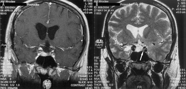
Figure 9.
(A) Post-contrast T1-weighted image, coronal plane: the residual tumour demonstrates enhancement pattern exactly the same as normal pituitary gland making correct diagnosis impossible. (B) Pre-contrast T2-weighted image, coronal plane: the intrasellar mass presents with high signal intensity, what indicates residual tumour and excludes recognition of the pituitary gland, which is visible on the left side of the sella (arrow).

