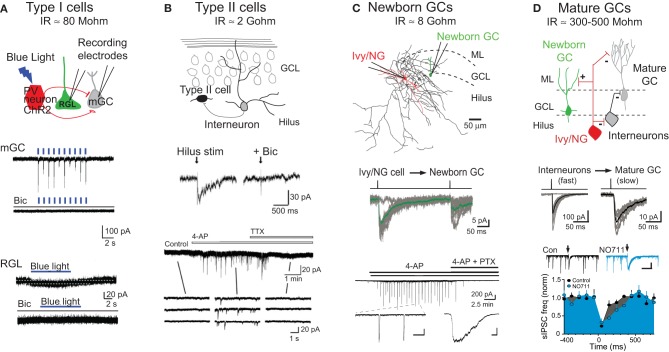
Figure 2.
Modes of GABAR signaling during GC development. (A) Tonic signaling to Type I radial glial-like stem cells: Top, schematic depiction of whole cell patch clamp recording from mature granule cells (mGCs) or Type I GFP+ radial glial-like stem cells (RGLs) during photoactivation of PV+ interneurons expressing channelrhodopsin (ChR2). Middle, photoactivation of ChR2+PV+ interneurons (indicated by blue lines, 1 Hz) generates large synaptic currents in mature GCs that are blocked by the GABAR antagonist bicuculline (Bic). Bottom, photoactivation of ChR2+PV+ interneurons (blue bar, 8 Hz) generates small tonic currents in Type I RGLs that are blocked by bicuculline. Adapted from Song et al. (2012) with permission from Macmillan Publishers Ltd. Estimates for input resistance (IR) of Type I and Type II cells are from Fukuda et al. (2003). (B) Synaptic signaling to Type II neuroprogenitors: Top, schematic depiction of innervation of Type II cells by dentate interneurons. Middle, hilar stimulation evokes inward currents in Type II cells that are blocked by bicuculline. Bottom, the frequency of spontaneous activity in Type II cells is increased by bath application of 4-aminopyridine (4-AP; 100 μM), a potassium channel blocker that causes synchronous firing of dentate interneurons (Michelson and Wong, 1994; Markwardt et al., 2011). 4-AP-induced currents are blocked by tetrodotoxin (TTX). Insets show currents on an expanded time scale. Adapted from Tozuka et al. (2005) with permission from Elsevier. (C) Synaptic signaling to newborn GCs by Ivy/NG interneurons: Top, reconstructed Ivy/NG interneuron (red cell body and dendrites, black axon) from a paired recording with a POMC-GFP+ newborn GC (green). ML, molecular layer. Middle, current injection into presynaptic Ivy/NG cells evokes slow GPSCs in newborn GCs. The averaged unitary IPSC (green) is overlaid on individual currents (gray). From Markwardt et al. (2011). Bottom, bath application of 4-AP (100 μM) induces synaptic currents in newborn GCs that are blocked by picrotoxin (PTX). Insets show currents on expanded time scales [scale bars are 200 pA and 10 s (left) or 500 ms (right)]. From Markwardt et al. (2009). The estimate for IR of newborn GCs is from Overstreet et al. (2004). (D) Heterogeneous synaptic signaling to mature GCs: Top, schematic depiction showing that mature GCs are innervated by a variety of interneuron subtypes including Ivy/NG cells and perisomatic projecting interneurons. Ivy/NG cells also provide slow GABAergic inhibition to perisomatic-projecting interneurons. Middle, current injection in individual interneurons evokes either fast (left) or slow (right) uIPSCs in mature GCs, reflecting the heterogeneity of innervation. Bottom, spontaneous GPSCs in mature GCs arise from perisomatic-projecting interneurons (Soltesz et al., 1995). The frequency of sGPSCs is reduced by stimulation of the ML (arrows) that evokes slow NO711-sensitive IPSCs in mature GCs (insets) and perisomatic projecting interneurons (not shown). Together these results support the circuit diagram shown above. Scale in inset, 300 pA, 500 ms. Adapted from Markwardt et al. (2011). The estimate of IR for mature GCs is from Schmidt-Hieber et al. (2004) and Overstreet et al. (2004).

