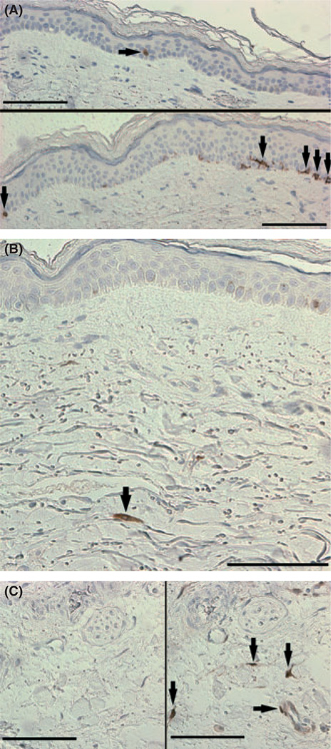
Fig. 1.
p16INK4a staining in human skin. (A) Representative p16INK4a staining of the epidermis from a subject in the middle tertile with one positive cell visible and an image of a section from a subject in the highest tertile (lower image with five positive cells visible). Epidermal staining was located along the basal membrane and mainly nuclear/perinuclear in nature although some cells displayed extensive cytoplasmic staining; note the dendritic nature of the fourth from right positive cell in the lower image characteristic of a melanocyte. (B) Representative p16INK4a staining of the dermis. (C) Negative control (no primary antibody, left image) and positive control (right image) of a skin sample used during all staining because of the consistent positive staining seen throughout the tissue. Line bars represent 100 µm (one dermal counting field was 315 by 315 µm) and black arrows the locations of positively stained cells.

