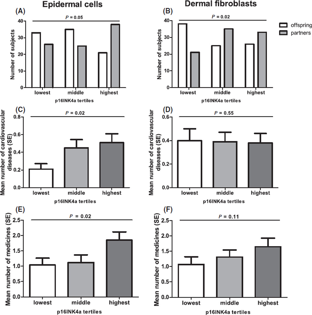
Fig. 2.
Tertiles of p16INK4a positivity in human skin. Left column (A, C, E) presents epidermal cells, the right column (B, D, F) presents dermal fibroblasts. (A, B) A comparison of distribution of tertiles of p16INK4a positive cells between offspring from long-lived families and their partners, N = 178. (C, D) Average number ± standard error (SE) of cardiovascular diseases over tertiles of p16INK4a positive cells, N = 155. (E, F) Average number ± SE of medicines over tertiles of p16INK4a positive cells, N = 136. Tertiles of p16INK4a positive epidermal cells: lowest ≤ 0.30 (median = 0.00), middle 0.30–1.30 (median = 0.55), highest ≥ 1.30 (median = 3.09) cells per mm length of the epidermal–dermal junction. Tertiles of p16INK4a positive dermal fibroblasts: lowest ≤ 0.72 (median = 0.00), middle 0.72–2.05 (median = 1.29), highest ≥ 2.05 (median 3.20) cells per 1 mm2 dermis. P-values are adjusted for age, gender, and smoking in (A) and (B); for gender and smoking in (C), (D), (E) and (F).

