
Figure 5.
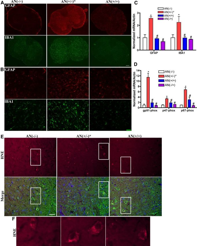
Overexpression of Nrf2 in astrocytes reduces gliosis and oxidative stress in spinal cord. A, Fluorescent staining of GFAP (red) and IBA1 (green); n = 3. Scale bar, 200 μm. B, Representative images of GFAP and IBA1 in ventral horn with higher magnification. Scale bar, 50 μm. C, mRNA levels of GFAP and IBA1 increased in spinal cord of 6-month-old hSYNA53T mice with symptoms [AN(+/−)*] compared with wild-type [AN(−/−)], double transgenic mice [AN(+/+)], and GFAP-Nrf2 littermates [AN(−/+)]. Mean ± SEM; n = 4. *p < 0.05, AN(+/−)* versus AN(−/−); #p < 0.05, AN(+/+), AN(−/+) versus AN(+/−)*. D, mRNA levels of NADPH oxidase subunits gp91-phox, p47-phox, and p67-phox increased in spinal cord of 6-month-old hSYNA53T mice with symptoms compared with wild-type and double transgenic mice. Mean ± SEM; n = 4. *p < 0.001, AN(+/−)* versus AN(−/−); #p < 0.01, AN(+/+), AN(−/+) versus AN(+/−)*. E, Intensity of HNE staining (red) increased in motor neurons in spinal cord of hSYNA53T mice. Green, β-III-tubulin; blue, Hoechst; n = 3. Scale bar, 50 μm. F, Higher magnification images of the square areas.