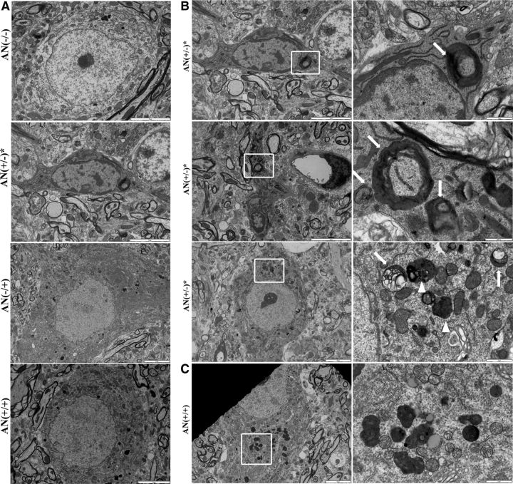
Figure 9.
Autophagosome-like vacuoles exist in hSYNA53T mice, not hSYNA53T/GFAP-Nrf2 double transgenic mice. A, Electron microscopic (EM) images of motor neurons in lumbar spinal cord from 8-month-old hSYNA53T mice, hSYNA53T/GFAP-Nrf2 mice, and littermate controls. B, Top and middle, autophagosome-like vacuoles in cell body and axon of neurons from hSYNA53T mouse with symptoms. Scale bars: 5 μm. Higher magnification images of the square areas are shown on the right. Scale bars: 1 μm (top) and 500 nm (middle). Arrows denote the vacuoles. Bottom, A healthier looking neuron from hSYNA53T mouse with vacuolated (arrows) and normal (arrowheads) lysosomes. Scale bars: 5 μm (left) and 1 μm (right). C, EM images for hSYNA53T/GFAP-Nrf2 mouse. Higher magnification images of the square areas are shown on the right. Scale bars: 5 μm (left) and 1 μm (right).

