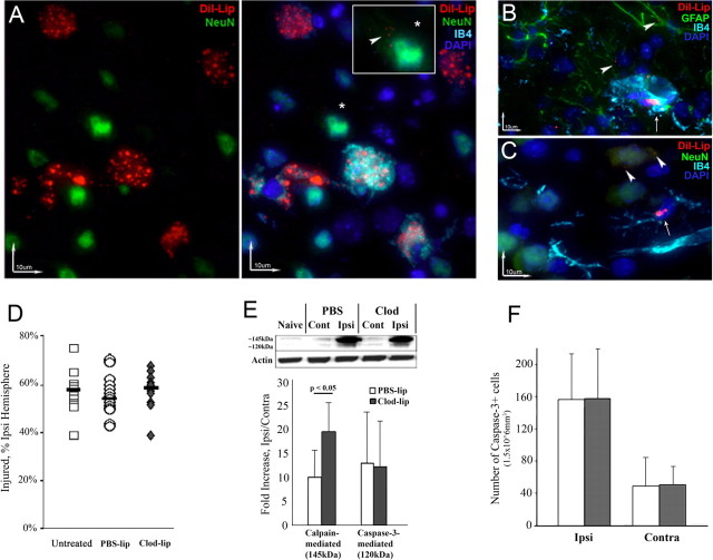
Figure 2.
Depletion of microglial cells does not affect volume of initial injury but shifts the balance between calpain- and caspase-3-dependent spectrin degradation 24 h after MCAO. A, Dil-lip predominantly accumulate in ameboid microglial cells in injured brain regions (3D reconstruction; red, Dil-lip; green, NeuN; blue, DAPI; turquoise, IB4). *Larger image (in the box) of a neuron with few small DiI-lip. Note the immensely different extent of DiI incorporation between neurons and microglia. B, C, The extent of DiI-lip incorporation in non-ameboid microglia is considerably lower. Only small Dil-lip occasionally accumulate in astrocytes (B, arrowhead) and neurons in uninjured tissue (C, arrowhead). Arrows, microglia. D, Volume of DWI-identifiable injury during MCAO is similar in untreated, PBS-lip and Clod-lip-treated animals. Shown are data for individual animals. Horizontal bars are medians. E, Clod-lip enhances calpain-mediated spectrin cleavage (the ∼145 kDa band) but does not affect caspase-3-mediated spectrin cleavage (the 120 kDa), n = 7–8 per group. F, The number of cells with cleaved caspase-3 in the penumbra is unchanged by Clod-lip treatment, n = 6–8 per group.

