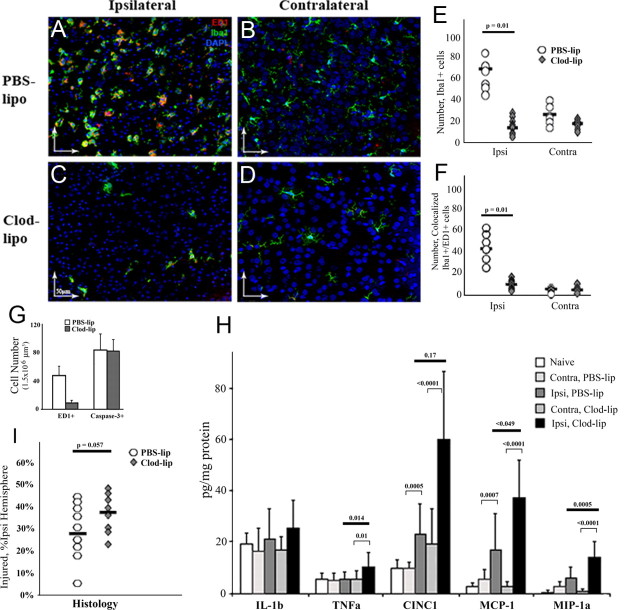
Figure 4.
Depletion of microglia increases cytokine and chemokine accumulation and exacerbates injury 72 h after reperfusion. A–D, In PBS-lip-treated pups, Iba1+ cells are abundant in the ischemic core (A, the activated phenotype) and in contralateral hemisphere (B, the resting phenotype). In Clod-lip-treated pups, the number of Iba1+ cells remains significantly lower than in vehicle-treated pups in both injured (C) and contralateral (D) tissue. Double-labeled Iba1+/ED1+ are predominantly seen in the ischemic core (A, C) and are rarely seen in contralateral hemisphere (B, D). Green, Iba1; red, ED1; blue, DAPI. E, F, The numbers of Iba1+ (E) and Iba1+/ED1+ cells (F) are significantly lower in injured tissue of Clod-lip-treated pups. The difference is less profound in the contralateral hemisphere, n = 8–9 per group. G, The absence of ED1+ cells does not affect the number of cells with cleaved caspase-3. Data shown as average ± SD. n = 8–10. H, The cytokine and chemokine levels (multiplex) in Clod-lip and PBS-lip-treated pups. n = 7–8 per group. I, Clod-lip treatment increases volume of histological injury in pups during subacute injury phase (Nissl staining), n = 10–13 per group. Black bars, Medians.

