
Figure 1.
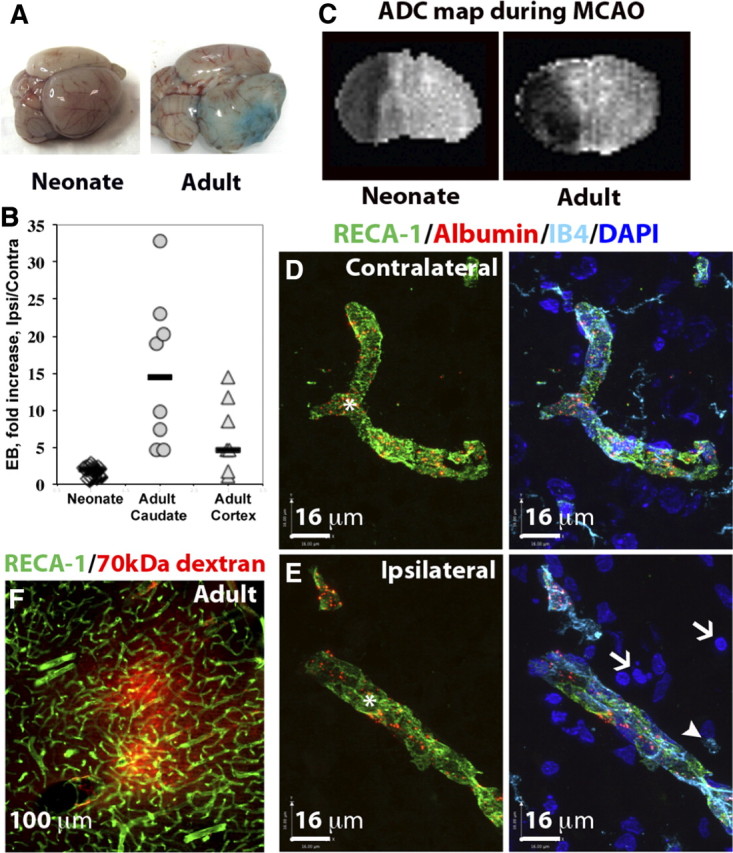
Blood–brain barrier permeability is markedly increased in injured brain regions in the adult but is largely preserved in the neonate after acute MCAO. A, Representative whole brains showing Evans blue extravasation and accumulation in neonatal and adult brain 24 h after reperfusion. B, Quantification of intravenously administered Evans blue in the brain between 2 and 24 h after reperfusion. Evans blue accumulation is profoundly increased in the adult but not in the neonatal rat. Numbers show how many times greater the accumulation of Evans blue occurred in the injured region as compared to the contralateral region in the same rat. Shown are data for individual rats; horizontal bars indicate medians. C, Apparent diffusion coefficient (ADC) maps showing a similar extension of brain edema during MCAO in neonate and adult brains. D, E, The spatial distribution of intravenously administered Alexa-555-conjugated albumin in contralateral (D) and injured (E) cortical regions in neonates 24 h after reperfusion. Injured areas in the ipsilateral hemisphere (E) were identified by the presence of pyknotic nuclei (DAPI, white arrows) and round, ameboid-like IB4+/RECA-1− microglia/macrophages (white arrowheads). In the injured areas, intravenous tracers colocalized with brain vessels (RECA1+/IB4+, asterisk) and were not observed in the extravascular spaces or in phagocytic microglia/macrophages (white arrowheads). Green, RECA-1; turquoise, IB4; blue, DAPI. Sections are 12 μm thick. F, Extravasation of Alexa-555-conjugated albumin into the injured cortex of adult rats 24 h after reperfusion (section thickness, 50 μm).