
Figure 7.
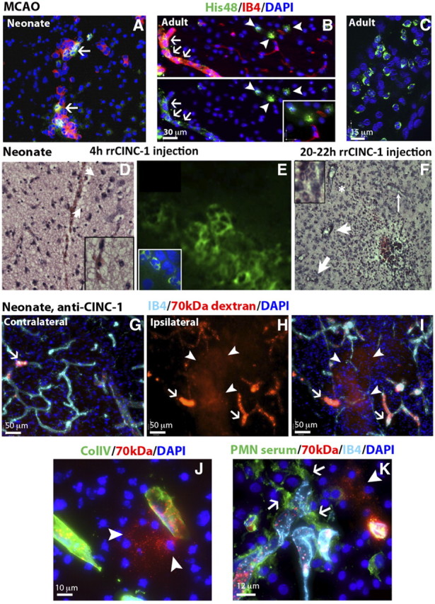
Neutrophil transmigration is limited after acute neonatal stroke. A, Neutrophil transmigration does not occur within 24 h after MCAO but His48+ cells are seen within vessels in animals without intracardiac perfusion (IB4, red; His48+ cells, green). B, C, Neutrophils (His48, green) are present in the brain parenchyma (B, arrowheads), meninges (C), and in a subpopulation of brain vessels (IB4, red) (B, arrows) in adult animals 4 h after reperfusion. D, E, Neutrophils are observed on H&E (D) and His48-immunofluorescence image (E) 4 h after intracerebral injection of rrCINC-1. Arrows in D point at neutrophils. His48+ cells are seen along the needle track and in parenchyma, but only a few His48+ neutrophils transmigrate following intracerebral rrCINC-1 injection (E). F, H&E staining showing infiltration of neutrophils in the brain 20–22 h after intracerebral injection of rrCINC-1. Neutrophils are observed in association with brain vessels (thin arrows) and in the brain parenchyma (thick arrows). Asterisk indicates the region magnified in the upper-left inset. G–I, Large areas of 70 kDa dextran (red) extravasation (H, I, arrowheads) were observed in the injured cortex of neonate rats injected with anti-CINC-1 15 min before MCAO. Increased accumulation of 70 kDa dextran (red) was also observed in brain vessels (arrows) in both the contralateral (G) and ipsilateral (H, I) cortex. (section thickness, 50 mm). J, Detail of a leaking vessel (70 kDa dextran, red, arrowheads) in a neonate rat injected with anti-CINC-1. K, Neutrophils (anti-PMN serum, green, arrows) associated with a leaking vessel (70 kDa dextran, red, arrowhead) in the injured cortex of a neonate rat injected with anti-CINC-1.