
Fig. 6.
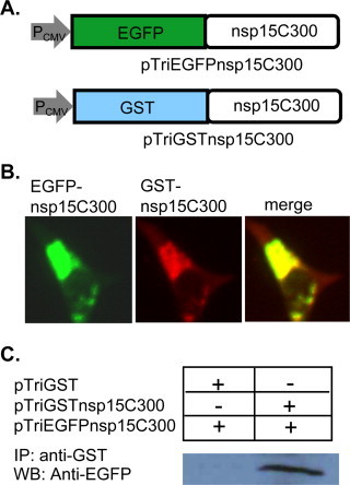
Protein-protein interaction involving the C-terminal domains of MHV nsp15. (A) Diagram of expression plasmids pTriEGFPnsp15C300 and pTriGSTnsp15C300 showing the MHV nsp15 C-terminal 74aa (from aa300 forward) coding sequence fused at the N-terminus to EGFP or GST, respectively. (B) Detection of co-localization (right panel, merge) of EGFP-nsp15C300 and GST-nsp15C300 by immunofluorescence staining with antibodies specific to GFP (left panel, green) and GST (middle panel, red) at 48 h post transfection. (C) Detection of the homotypic interaction involving the C-terminal domain of MHV nsp15 by co-immunoprecipitation and Western blot analysis. Cells were co-transfected with pTriEGFPnsp15C300 and pTriGSTnsp15C300 or pTriGST. Cell lysates were then precipitated with an antibody specific to GST. The immunocomplex was then detected by Western blot with an antibody specific to GFP. (For interpretation of the references to color in this figure legend, the reader is referred to the web version of the article.)