
Figure 1.
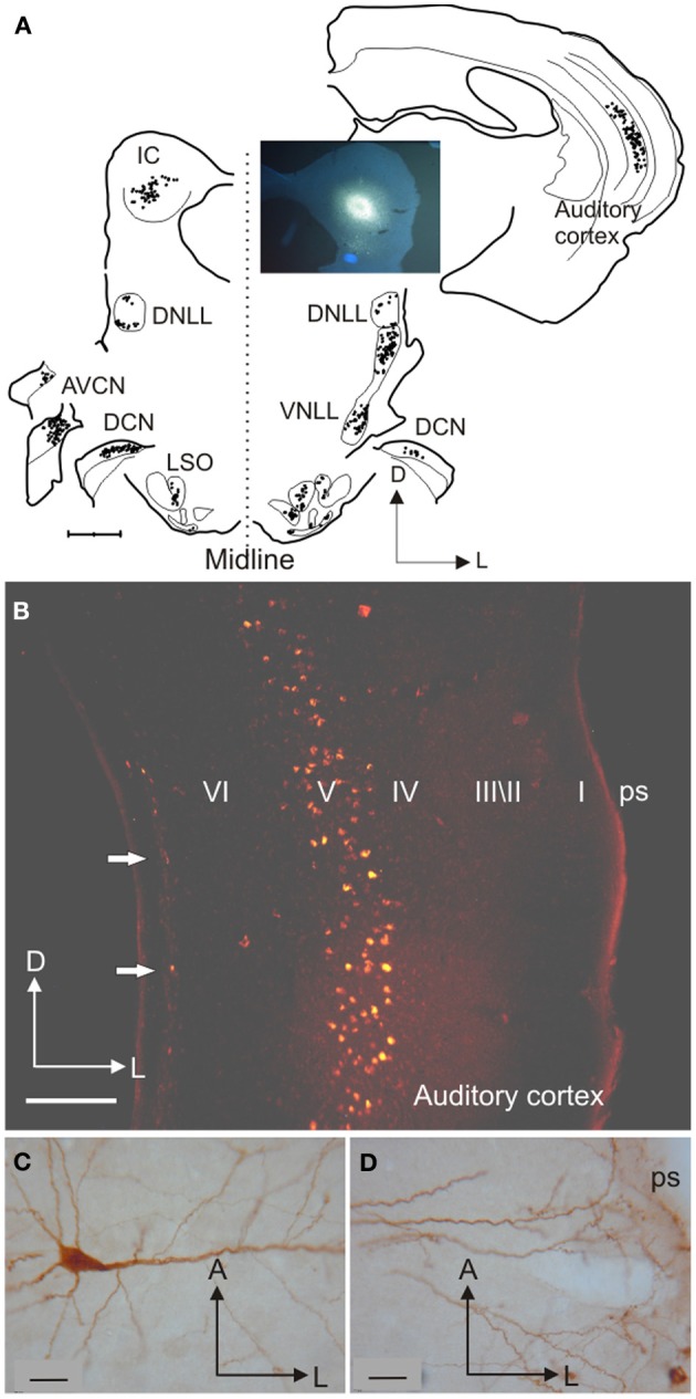
Inputs from different auditory centers converge in the IC. (A) A small fluorogold tracer injection in the ventromedial part of the IC central nucleus of the gerbil produces retrograde labeling of neurons in the MSO, periolivary nuclei, VNLL, and A1 on the same side, in the cochlear nuclei and IC on the opposite side, and in the LSO, DCN, and DNLL on both sides. (B) Retrogradely labeled cells in the cortex are found mainly in layer V after a rhodamine tracer injection in the IC. (C) Large labeled pyramidal cell with the soma located in cortical layer V and (D) a tufted dendritic tree ending in layer I. Calibration bars: 1 mm (A), 0.2 mm (B), and 25 μm (C,D). Modified with permission from Bajo and Moore (2005).