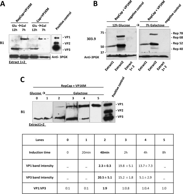
Figure 4.
VP1-VP3 expression pattern in co-transformed yeast clones. Cells co-transformed with YEplacp40Cap and pYESVP1KM (Cap + VP1KM clone) (A) and Rep expressing cells co-transformed with YEplacRepCap and pYESVP1KM (RepCap + VP1KM) (B), were grown on glucose and then transferred to galactose for induction. Equal amounts of total cellular proteins (extracts 1 + 2) were analyzed by Western blot, using mAb B1 to detect VP proteins. (A): VP3 was detected in both yeast clones after 12 h growth in glucose and it decreased along with VP1 induction upon 7 h in galactose. (B): Extracts from RepCap + VP1KM clones were analyzed for Rep protein expression before (12 h glucose) and after 7 h galactose growth, using mAb 303.9. Similar amounts and distribution of Rep isoforms to extracts 1/2 were detected in glucose and galactose samples. Extracts from cells co-transformed with empty vectors, YEplac181 and pYES2 were used as -control. (C): Lanes 0–5: VP1-VP3 expression pattern in total cell-extracts derived from RepCap + VP1KM clones before induction (“0” time point) and at various times of galactose induction. VP1:VP3 ratios are determined band densitometry and shown in the table below. Numbers represent the density expressed in arbitrary unit detected by the analysis software described in materials and methods. Results are reported as mean of at least three independent experiments ± standard error. The best ratio was obtained after 40 minutes of galactose induction.

