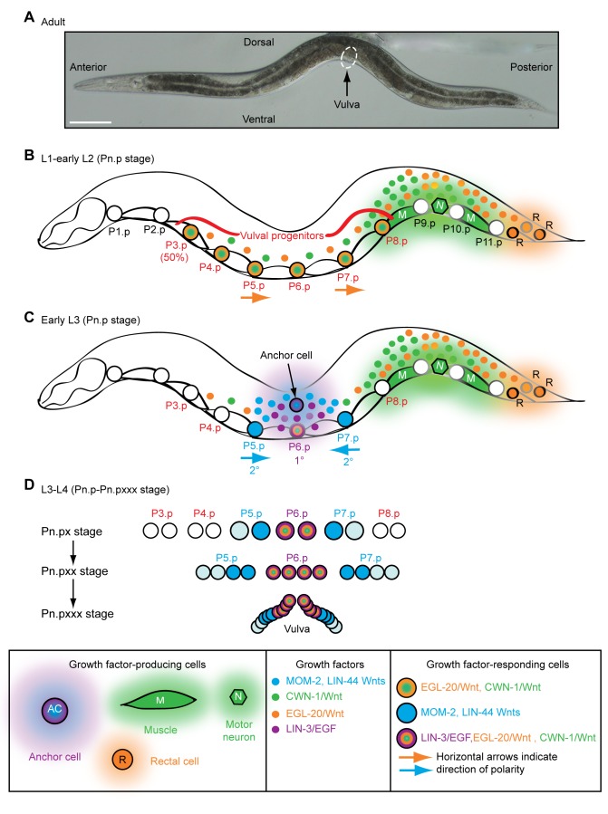
Figure 1. Wnt signaling and epidermal patterning in C. elegans.
(A) A wild-type C. elegans adult hermaphrodite. Scale bar is 100 µm. (B) During the L2 larval stage, LIN-3/EGF from pre-anchor cell/ventral uterine precursor cells (not shown) cooperates with a gradient of EGL-20/Wnt (orange) from rectal cells and CWN-1/Wnt (green) from posterior muscle and neurons to cause six epidermal cells to become vulval progenitors (P3.p–P8.p). 50% of the time, P3.p does not receive sufficient Wnt signaling and adopts the “F” fate (also known as the 4° fate) and fuses with a hypodermal syncytium called hyp7. EGL-20/Wnt also polarizes P5.p and P7.p so that they face posteriorly (horizontal arrows). The epidermal cells normally touch each other, but are drawn apart to facilitate depiction of muscle and neurons. (C) At the end of the L2 larval stage, anchor cell-produced MOM-2 and LIN-44 Wnts (blue) reorient P7.p towards the anterior (horizontal arrows). During the L3 larval stage, LIN-3/EGF (purple) from the anchor cell induces the 1° vulval fate in P6.p, which is facilitated by EGL-20 and CWN-1 Wnts. P5.p and P7.p adopt 2° vulval fates because of the activation of LIN-12/Notch via a lateral signal from P6.p. (D) During the L3–L4 larval stages, vulval progenitor cells (Pn.p) divide to generate Pn.px cells, with P5.p–P7.p undergoing two additional rounds of cell division (to ultimately make Pn.pxxx cells). Because of the opposite polarities of P5.p and P7.p, their asymmetrically dividing progeny generate mirror image patterns. By the early L4 stage, a 22-cell vulva is generated. The Pn.px progeny of P3.p, P4.p, and P8.p fuse with hyp7 (3° fate).

