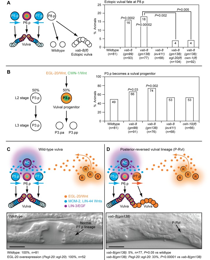
Figure 2. vab-8 mutations that affect neuronal cell body positioning and axon outgrowth cause epidermal patterning defects.
(A) vab-8 mutations cause P8.p to adopt a vulval fate. In wild-type animals, P8.p divides once, but never forms vulval tissue. (B) vab-8 mutations increase the frequency of P3.p becoming a vulval progenitor. 50% of the time, P3.p receives sufficient Wnt signaling to become a vulval progenitor and divides once. (C) Upper panel depicts wild-type signaling by Wnts and EGF that promotes vulval development with mirror image symmetry. MOM-2 and LIN-44 Wnts dominate over EGL-20/Wnt to polarize P7.p towards the anterior. Lower panel shows a wild-type 22-cell vulva with normal symmetry at the mid-L4 stage. (D) Upper panel depicts abnormal Wnt signaling in vab-8 mutants that causes the formation of vulval tissue with a P-Rvl phenotype. EGL-20/Wnt dominates over MOM-2 and LIN-44 Wnts, preventing P7.p from reorienting towards the anterior. Lower panel shows a P-Rvl vulva at the mid-L4 stage. In (C) and (D), EGL-20/Wnt was overexpressed from its native promoter with the muIs49 transgene. Scale bar is 10 µm. Colors depict Wnt signaling as in Figure 1. p-Values were calculated using a two-tailed Fisher's exact test versus wild-type animals (A and B) or as otherwise indicated (A and D).

