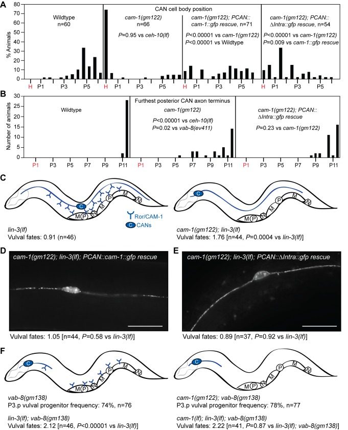
Figure 7. The CANs use the extracellular Wnt-binding domain of Ror/CAM-1 to direct epidermal patterning.
(A and B) Distributions of positions of CAN cell bodies and furthest posterior CAN axon termini in ror/cam-1 mutants and in cam-1 mutants expressing wild-type or intracellular-domain-deleted CAM-1(ΔIntra) only in the CANs. In non-ror/cam-1 rescue experiments, CANs were visualized with the kyIs4[Pceh-23::gfp] transgene. In ror/cam-1 rescue experiments, CANs were visualized by expression of GFP-tagged Ror/CAM-1 in the CANs. In ror/cam-1 rescue experiments, strains also harbored an egf/lin-3(lf) mutation. x-Axis indicates Pn.p or Pn.px positions. H, head/pharyngeal region. p-Values were calculated using a two-tailed Mann-Whitney U test. (C) Ror/CAM-1 inhibits vulval fate signaling in central vulval progenitors. (D) Transgenic CAN-specific expression of Ror/CAM-1::GFP restores inhibition of vulval development in ror/cam-1 mutants. (E) Transgenic CAN-specific expression of a Ror/CAM-1::GFP mutant lacking the intracellular domain (ΔIntra, see Figure 6C) also restores inhibition of vulval development in ror/cam-1 mutants. In (D) and (E), scale bar is 20 µm. (F) Under physiologic conditions, the majority of Ror/CAM-1 inhibition of vulval fate signaling is mediated by the CANs. If the CAN cell bodies are anteriorly displaced and the posterior axons are severely foreshortened, loss of Ror/CAM-1 activity from all cells does not further increase P3.p progenitor frequency or the amount of vulval development in sensitized backgrounds. Drawings depict the cellular distribution of Ror/CAM-1 as described in Figure 6E. M, muscle cells; N, neurons; P, vulval progenitors. In (C–E), vulval fates: number of vulval progenitor cells adopting vulval fates. Wild-type is 3.00. p-Values were calculated using a two-tailed Student's t test. The PCAN::cam-1::gfp and PCAN::ΔIntra::gfp rescuing transgenic arrays are dyEx44 and dyEx45, respectively.

