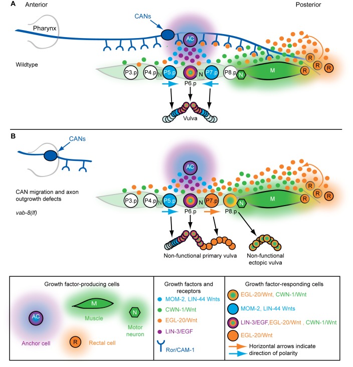
Figure 9. Model for neuronal control of axial patterning by Wnts.
(A) In wild-type animals, EGL-20/Wnt (orange) (and possibly CWN-1/Wnt [green]) is sequestered by the Ror/CAM-expressing posterior CAN axons. At the end of the L2 larval stage, this sequestration allows MOM-2 and LIN-44 Wnts (blue) to reorient and polarize P7.p towards the anterior (horizontal arrows). During the L3 larval stage, anterior-facing P7.p divides with the mirror image pattern of P5.p, and P8.p does not receive sufficient Wnt or EGF signaling to adopt a vulval fate. (B) If the posterior CAN axons do not extend far enough into the posterior body, where EGL-20/Wnt is produced, an abnormally high concentration of EGL-20 occurs, which prevents anterior polarization of P7.p, and causes ectopic vulval tissue to form at P8.p.

