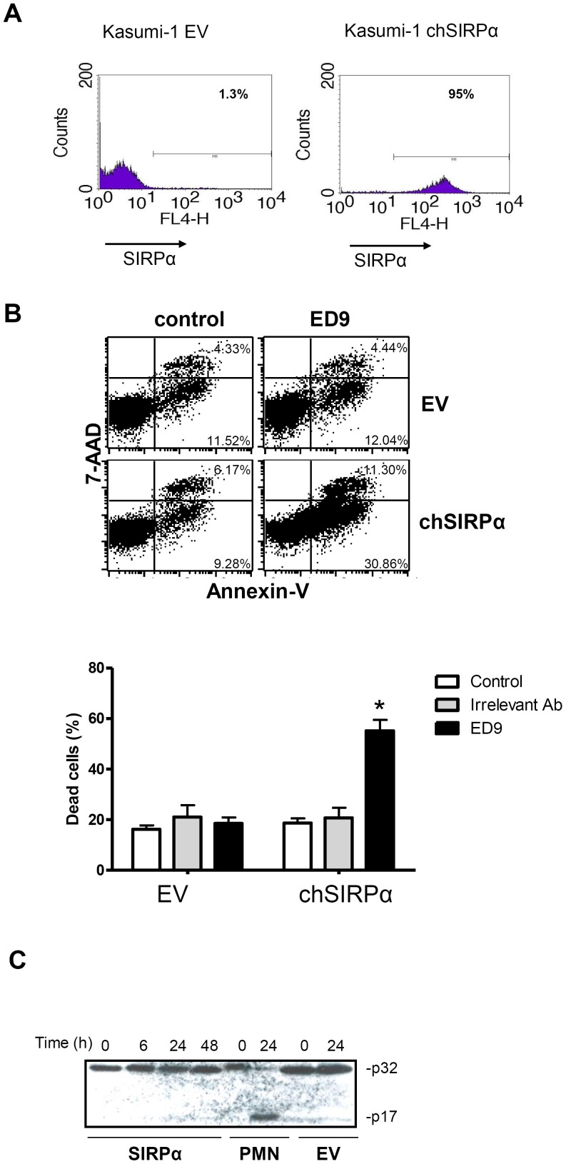
Figure 5. Ligation of chSIRPα induces caspase 3-independent PCD in Kasumi-1 cells.
(A) Flow cytometric analysis of SIRPα expression was performed by using ED9 mAb in stable Kasumi-1 cells expressing chSIRPα and EV. (B) Kasumi-1 chSIRPα and EV cells were incubated with 10 µg/ml ED9 mAb and the percentage of cell death was determined after 24 hrs. Annexin V and 7-AAD FACS staining defined that ligation of chSIRPα resulted in increased cell death in chSIRPα Kasumi-1 cells compared to EV control cells. Data are means ± SD calculated from 3 independent experiments using triplicate samples (*: significant difference p<0.05). (C) Kasumi-1 cells expressing chSIRPα or EV were treated with 10 µg/ml ED9 for mentioned time points. Caspase 3 staining shows no cleavage of the p32 subunit. As a positive control for caspase 3 cleavage, human neutrophils (PMN) were incubated at room temperature for 0 and 24 hours.

