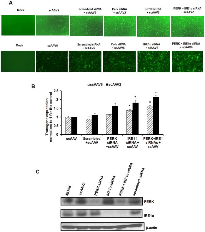
Figure 5. Comparative analysis of AAV mediated transduction efficiency in HeLa cells after siRNA mediated knock down of PERK or IRE1α pathways.
A. Transgene expression was measured in HeLa cells 48 hrs post-infection with self-complementary AAV2-EGFP or AAV6-EFGP vectors either in the presence or absence of specific siRNA or scrambled siRNA control. B. Quantitative analyses of the data from (A) by fluorescence microscopy. Images from five visual fields were analyzed quantitatively by ImageJ analysis software. Transgene expression was assessed as total area of green fluorescence (pixel2) per visual field (mean ± SD) and normalized to 1 for the control. Error bars represent standard error and the graph is a representative data set of at least three independent experiments. *p<0.05 Vs scrambled siRNA treated cells C. Western blot analysis of HeLa cellular extracts following mock (PBS)-infection or infection with AAV vectors, either in the presence or absence of PERK or IRE1α siRNA or scrambled siRNA control. β-actin was used as a loading control.

