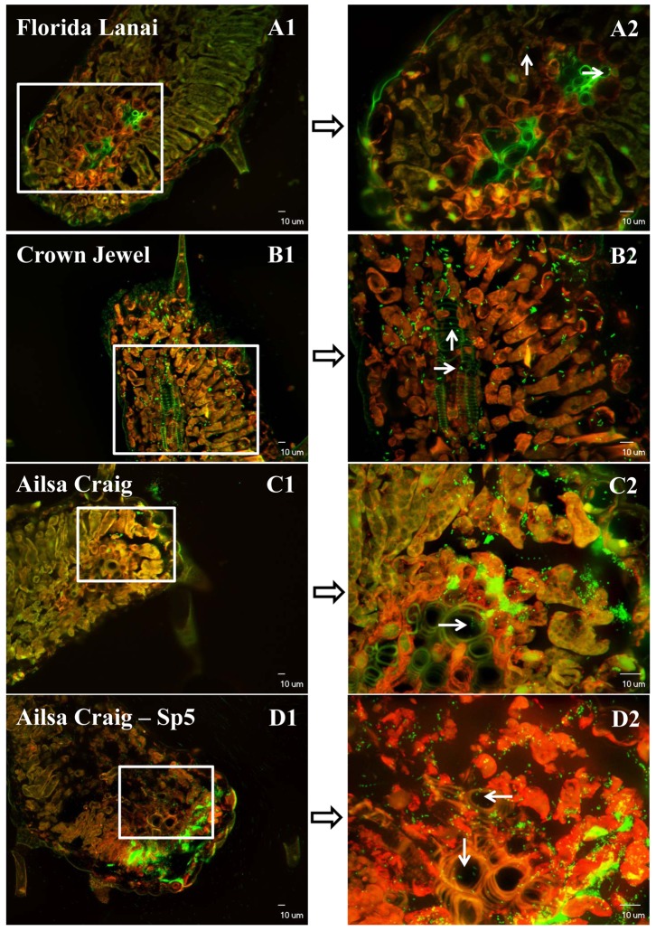
Figure 3. Confocal laser microscope images of tomato leaf tissue sections colonized by Salmonella Typhimurium after inoculation (109 CFU/ml) through guttation droplets.
White arrows point out the GFP-tagged Salmonella cells (green) which entered into the vascular system of tomato leaves. Red fluorescence is the autofluorescence of plant chloroplasts. Images A2, B2, C2 and D2 are merged images under GFP and TRITC filters obtained by projecting 20 Z section overlaid fluorescence images of different layers with 1 µm interval into one combined image [11].

