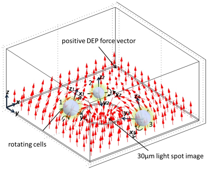
Figure 5. Illustration of cell rotation in an OEK.

The cells in the dark-field region rotated toward the 30 µm spot image. Their z-axes were normal to the positive DEP force vectors. The axis of rotation was at the x-axis perpendicular to the E-field.
