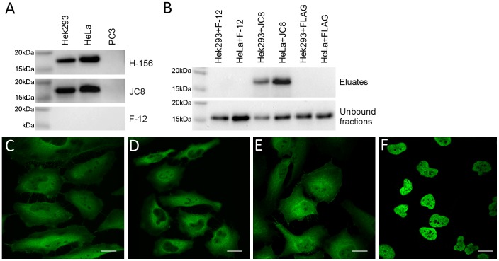
Figure 1. P16INK4A antibody validation.
P16INK4A subcellular localization by immunofluorescence in HeLa cells using anti-p16INK4a antibodies F-12 (A), H-156 (B), JC8 (C), and E6H4 (D); magnification bar = 20 µm. E. Comparison of anti-p16INK4a antibodies in Western Blot analysis using p16INK4a positive Hek293 and HeLa whole cell lysates and p16INK4a negative PC3 cell extract (each loaded 20 µg whole protein per well). F. Immunoprecipitation of p16INK4a using F-12 and JC8 mouse monoclonal antibodies, the FLAG mouse monoclonal antibody was used alongside as a negative control.

