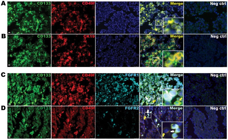
Figure 1. The murine E12.5 liver is populated with CD133posCD49fpos hepatoblast progenitor cells expressing FGFR1 and 2.
(A) Immunofluorescence staining for progenitor cell markers CD133 and CD49f on E12.5 embryonic liver. Yellow cells are double positive cells. DAPI staining (blue) identifies nuclei. (B) Immunostaining of CD133 and Cytokeratin (CK19). (C) Immunostaining for CD133, CD49f and FGFR 1 and (D) FGFR2. White arrows mark triple positive cells and yellow arrows mark positive cells that do not express receptors. Data are representative of three or more independent experiments. Scale bar represents 25 µm.

