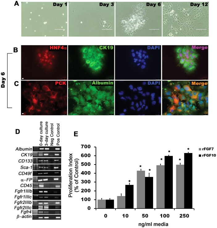
Figure 2. Isolation and in vitro expansion of CD133posCD49fposCD45neg cells by Magnetic Assisted Cells Separation (MACS) from E12.5 liver enriches for hepatoblast progenitor cells that proliferate in response to FGFR activation.
(A) Phase contrast pictures of in vitro expanded CD133pos CD49fpos CD45neg enriched cells from E12.5 embryonic liver at different time points. Co-immunofluorescence staining for (B) HNF4α and CK19 and (C) ALBUMIN and PCK. (D) Gene expression analysis by RTPCR using RNA isolated from the MACS enriched cells before and after 3-day culture. Negative control = water and positive control = E16.5 whole embryonic cDNA. Scale bar 25 µm. (E) Proliferation indices of CD133pos CD49fpos CD45neg cells treated with rFGF7/10 for 48 hrs and pulse labeled with BrdU (n = 4, *p<0.001 compared to control). Data are representative of three or more independent experiments.

