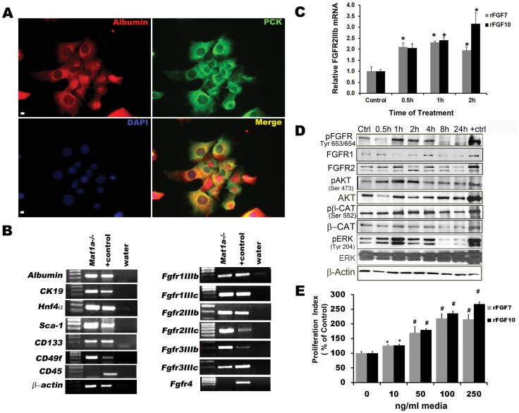
Figure 3. FGFR activation promotes downstream activation of AKT, ERK, and β-catenin pathways as well as proliferation of Mat1a .
−/− cells. (A) Co-localization of ALBUMIN and pCK in Mat1a−/− cells. Scale bar 25 µm. (B) RTPCR analysis shows Mat1a−/− cells express putative stem gene markers and Fgfr’s. Positive (+) control E16.5 = embryonic cDNA. Data are representative of three or more independent experiments. (C) qPCR analysis shows that rFGF7/10 upregulates expression of Fgfr2IIIb (n = 3, *p<0.01). (D) Western blot analysis of time course rFGF7 treatment on Mat1a−/− cells results in the downstream phosphorylation and activation of FGF receptor, AKT, ERK and β-CATENIN (ctrl (control) = untreated cells, +ctrl = E12.5 embryonic lung). Data are representative of two independent experiments. (E) Proliferation indices for Mat1a−/− cells treated with rFGF7/10 for 48 hrs and pulse labeled with BrdU (n = 4, *p<0.001 compared to control).

