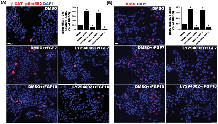
Figure 5. FGFR mediated proliferation of Mat1a .
−/− cells are mediated in part through PI3K-AKT pathway. Serum-starved Mat1a−/− cells were treated with rFGF7/10 ± PI3K-AKT inhibitor LY294002 for detection and quantification of (A) pSer 552 β-CATENIN or (B) BrdU. Total and β-CATENIN/BrdU Positive cells were counted from 4–5 HPF images from 3 independent experiments and represented as relative % of control (n = 3, *p<0.001).

