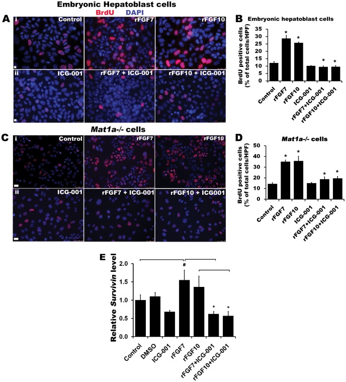
Figure 7. Activation of FGF signaling drives embryonic hepatoblasts and Mat1a −/− cells into the cell cycle through a CBP-dependent pathway.
Immunodetection and quantification of BrdU positive cells in (A, B) CD133pos CD49fpos Cd45neg enriched embryonic hepatoblasts (3-day cultured) and (C, D) Mat1a−/− cells after treating with rFGF7 or rFGF10 ± β-catenin-CBP inhibitor ICG-001. Total number of BrdU positive cells were counted from 3–4 HPF from three independent experiments and represented as % of total cells (n = 3, *p<0.0001 versus control). FGF-ICG-001 was compared to respective control FGF treated experiments. (E) qPCR analysis of Survivin mRNA expression level in Mat1a−/− cells for control, DMSO control, and rFGF7/10 ± ICG-001 groups. Expression levels are normalized to Actin (n = 3, *p< 0.005, #p<0.05).

