
Figure 3. Cytotoxic effect of BBE in oral epithelial (OKF6) cells.
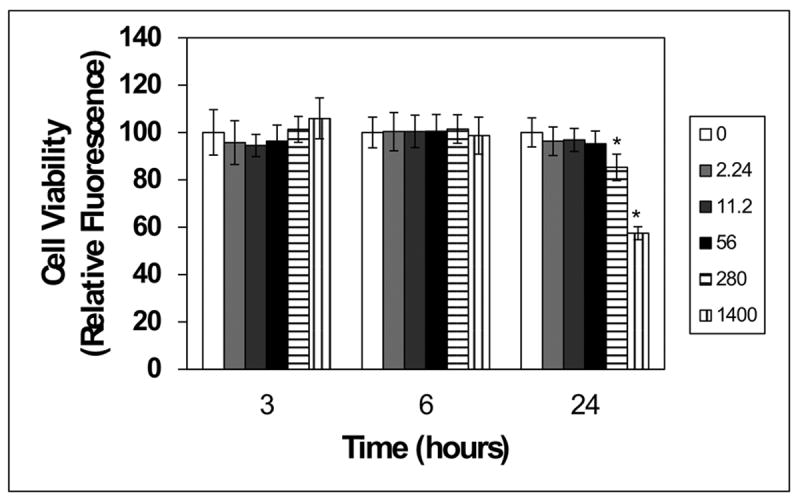
OKF6 cells were plated in 96-well plates (9,400 cells/well) in Ker-SFM for 24 h as described in the methods. Cells were treated with vehicle control (water/white bar) or varying concentrations of BBE for 3, 6 or 24 hours. Cell viability was evaluated using the CellTiter-Blue viability assay. Data for BBE-treated cultures were normalized to the mean for water-treated cultures. Values significantly different (p ≤ 0.05) between water-treated cells vs. OKF6 cells exposed to BBE are identified with an asterisk (*).