
Figure 1.
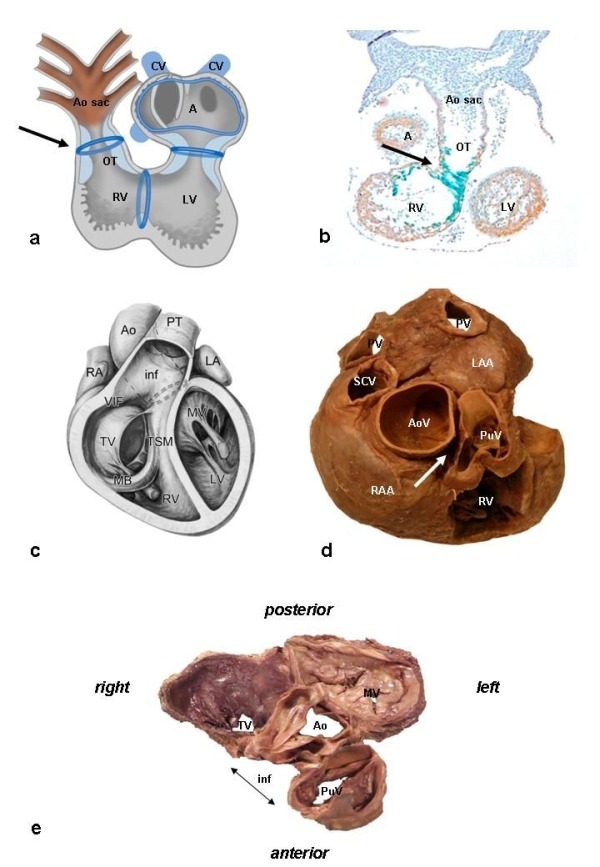
Embryology and Anatomy. Panel A: Schematic depiction of the heart tube during an early developing stage, after looping of the heart has been initiated. Several transitional zones, located in between the different cardiac segments, can be recognized in the heart based on histological characteristics as well as expression of molecular markers. These zones, indicated schematically in blue, will contribute to elements of the cardiac conduction system. The ventriculo-arterial transition, indicated by the arrow, is situated at the level of the outflow tract (OT) of the heart. In the adult heart, only part of these transitional zones can still be recognized, in elements of the definitive cardiac conduction system (not shown) ( reproduced with permission from Gittenberger-de Groot AC et al. James H. Moller , Julien I. E. Hoffman, eds. Pediatric Cardiovascular Medicine. Second ed.December 2011, Wiley-Blackwell). Panel B: Expression of the marker CCS-lacZ that can be found during embryonic heart development in elements of the cardiac conduction system and the transitional zones. Shown here is CCS-lacZ expression at the level of the OT (arrow) (reproduced with permission from Jongbloed et al. J Cardiovasc Electrophysiol 2004;15(3):349-355). Panel C: Anatomy of the outflow tract, anterior view, demonstrating the position of the great arteries in the normal heart. The aorta (Ao) is positioned right posteriorly in relation to the pulmonary trunk (PT). On the right side, the fibrous tissue of the tricuspid valve (TV) is separated from the fibrous tissue of the pulmonary valve by the posterior wall of the muscular infundibulum (inf) (reproduced with permission from Bartelings MM and Gittenberger-de Groot AC (1989) The outflow tract of the heart - embryologic and morphologic correlations. Int.J.Cardiol. 22, 289-300). Panel D: Superior view. The aorta has a central position and is wedged in between the atrioventricular orifices and appendages of the right atrium (RAA) and left atrium (LAA). The aortic valve (AoV) has an inferior position as compared to the pulmonary valve (PuV). Note that there is no continuity between the distal medial and posterior wall of the RVOT and the aorta (arrow). Panel E: The fibrous heart skeleton. Note the fibrous continuity between the aortic valve, mitral valve (MV) and tricuspid valve, whereas there is no continuity with the fibrous tissue of the pulmonary valve due the presence of the subpulmonary infundibulum. Other abbreviations: A: common atrium, AoS: aortic sac, CV: cardinal veins, LA: left atrium, LV: left ventricle, MB: moderator band, PV: pulmonary vein, RA: right atrium, RV right ventricle, SCV: superior caval vein, TSM: trabecula-septomarginalis.