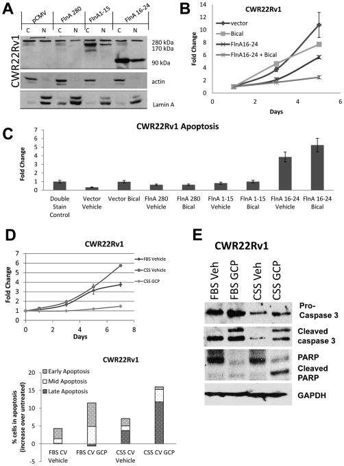
Figure 2. Treatment with genistein combined polysaccharide (GCP) replicates the effects of FlnA nuclear localization.
(A) Expression of the transfected proteins when CWR22Rv1 cells were transiently transfected for 48 hours with 2 μg/ml of an empty vector, full-length FlnA (280 kDa), FlnA 1-15 (170 kDa) or FlnA 16-24 (90 kDa). Actin and Lamin A were used as markers of the cytoplasm and the nucleus, respectively. (B) As estimated by MTT assay, neither 10 μM bicalutamide nor FlnA 16-24 individually had significant effects on the growth of CWR22Rv1 cells, but in combination, they prevented cell growth. Each point on the graph represents mean ± S.D. of 3 independent readings. (C) CWR22Rv1 cells underwent apoptosis when transfected with FlnA 16-24 but not when transfected with an empty vector, FlnA 280 or FlnA 1-15 as determined by flow cytometry after 48 hours in Annexin V and propidium iodide stained cells. (D) (upper) MTT assay showing effect of CWR22Rv1 cells with GCP or vehicle in FBS and CSS containing medium. Each point on the graph represents mean ± S.D. of 3 independent readings. (lower) Analysis of apoptosis by flow cytometry in propidium iodide and Annexin V stained cells demonstrating that CWR22Rv1 cells treated with 100 μg/ml GCP for 48 hours experienced more apoptosis than vehicle treated cells. Early apoptosis: Annexin V staining, Mid-apoptosis: Annexin V+PI staining, Late apoptosis: PI staining. Results presented represent numbers after subtraction from untreated cells. Note the increase in apoptosis in cells cultured in CSS vs FBS containing medium. (E) Western blots demonstrating that 100 μg/ml GCP after 48 hrs increased apoptosis as demonstrated by cleaved Caspase 3 and PARP levels.

