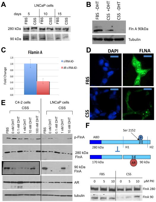
Figure 6. ADT prevents Filamin A cleavage to the 90 kDa fragment and phosphorylates FlnA at Ser 2152.
(A) LNCaP cells were cultured for up to 15 days in FBS or CSS containing medium, and the levels of 90 and 280 kDa FlnA were determined by western blotting. (B) Western blot showing decreased expression of the 90 kDa FlnA fragment in LNCaP cells in CSS vs FBS, whereas its levels were restored by the addition of 1 nM DHT. (C) FlnA expression in CWR22Rv1 cells is regulated by the AR. AR siRNA decreased FlnA mRNA expression by ∼60% as determined by qPCR in medium with CSS (AD-). (D) LNCaP cells were grown in either FBS or CSS containing medium, and FlnA protein expression was determined by immunofluorescence. Scale bar: 10 μm. Note that cells grown in FBS containing medium show higher expression of FlnA compared to cells grown in CSS containing medium. (E) LNCaP and C4-2 cells were cultured in FBS or CSS with increasing levels of DHT as indicated for 48 hrs. Western blots demonstrate the levels of expression of various proteins. Note that C4-2 cells actually have much lower levels of FlnA 90 kDa, therefore the blot shown here for C4-2 was at a higher exposure. (F) (Upper) Scheme showing FlnA cleavage at H1 is regulated by phosphorylation at Ser 2152. When FlnA is phosphorylated, it does not undergo cleavage and remains in the cytoplasm, whereas upon dephosphorylation, FlnA is cleaved to the 90 kDa fragment which translocates to the nucleus. (Lower) LNCaP cells were cultured in FBS or CSS with increasing levels of the PKA inhibitor PKI 14-22 (PKI) for 48 hrs. Western blotting demonstrated that culture in CSS depleted levels of the 90 kDa FlnA, but treatment with increasing doses of PKA inhibitor in CSS containing medium restored its expression.

